

Loyal companions
A pair of University of Minnesota doctors teams up to improve treatments for dogs and people facing brain cancer

Brain size varies by species. The brain of a hamster weighs about 1 gram. A dog’s brain is typically 5 centimeters in diameter, while the human brain is about three times as large.
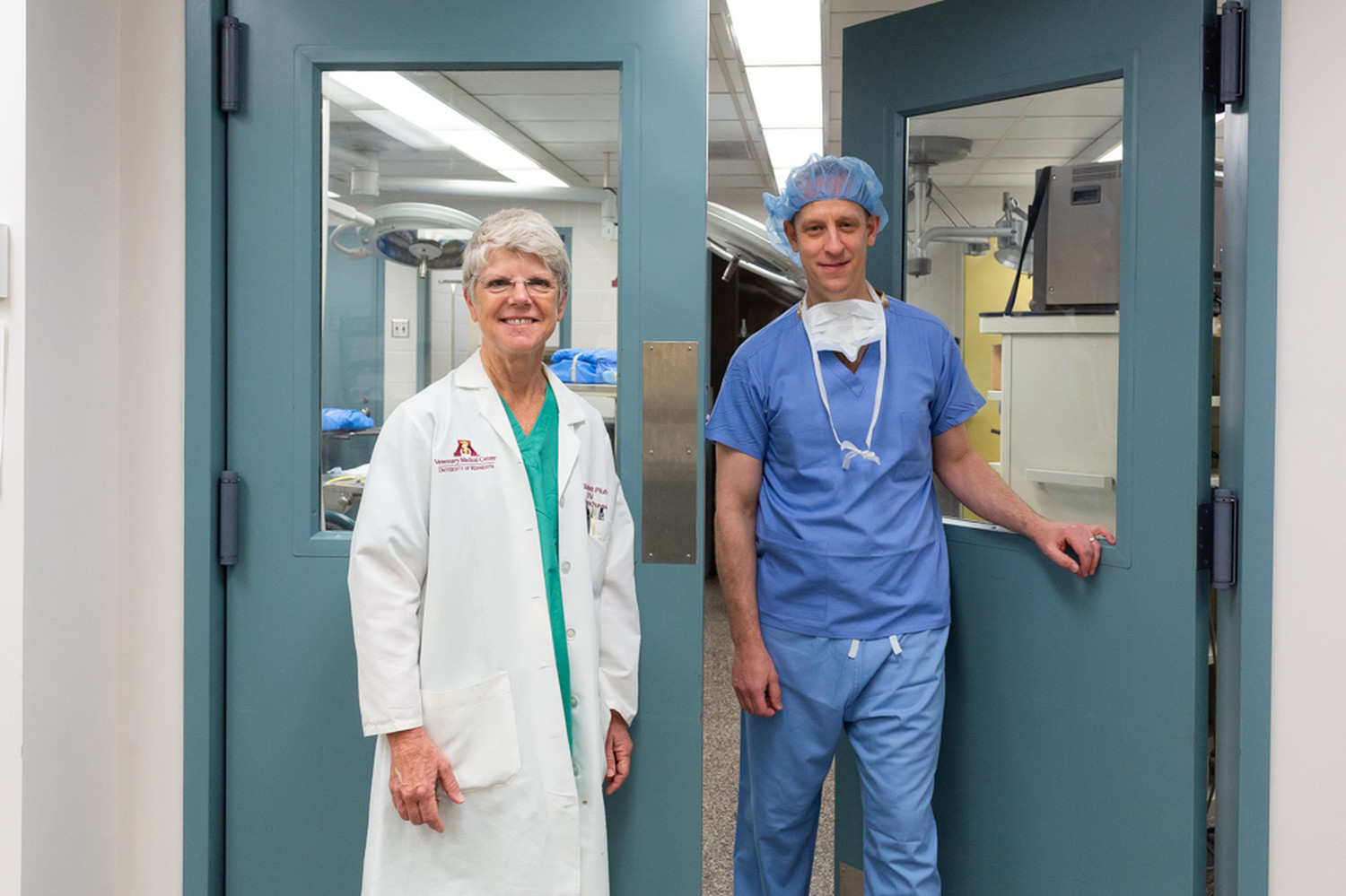
Despite such variations in size, however, the structure of brains across all mammalian species is remarkably the same. Those similarities are the basis for an ongoing collaboration between Elizabeth Pluhar, D.V.M., Ph.D., a professor in the University of Minnesota’s College of Veterinary Medicine, and Matthew Hunt, M.D., a neurosurgeon and associate professor of neurosurgery in the U’s Medical School.
Pluhar typically operates on dogs, while Hunt primarily treats human patients. But two or three days each month, the pair meets at the Veterinary Medical Center to perform surgery on dogs that have glioblastoma, a deadly form of brain cancer.
Their collaboration, part of a research initiative begun by the late U of M professor John Ohlfest, Ph.D., aims to find better ways to treat glioblastoma in humans while simultaneously offering relief to canines and bringing hope to their owners. It’s called comparative oncology, and the U is one of the only centers in the country with the expertise to do this work.
“We do more neurosurgery on dogs than any other veterinary center in the world,” Pluhar says of the unique collaboration. To date, she and Hunt—both members of the Masonic Cancer Center, University of Minnesota—have removed brain tumors from more than 200 canines.

Many similarities
For decades, brain cancer researchers tested treatments on mice and rats. If efforts to prevent, slow, or cure tumor development in rodents were successful, the theory went, perhaps the same thing would work in humans.
But in recent years, studies have suggested that naturally occurring cancers and artificially induced cancers respond to treatment in different ways.
Researchers needed to look beyond mice. The search for a better animal model eventually led to dogs.
“It just so happens that no other species spontaneously develops brain tumors at the rate or incidence that dogs do,” Pluhar says. “And the incidence in dogs is very similar to what it is in people.”
In shape and consistency, too, the glioma that occurs in dogs is almost identical to the one that can form in human brains.
In hopes of fighting cancer in both dogs and humans, Pluhar and Hunt are fusing their expertise. By integrating their experience in neurosurgery and veterinary care, they hope to accomplish two things: to improve cancer treatment for dogs, boosting long-term survival rates and preserving quality of life, and to use that information to better treat human brain cancers.
Tracking long-term success
Hunt, who joined the research effort in 2008, says operating on dogs is quite similar to working on humans.
“Things are named a little differently, but otherwise the brain anatomy and structure are largely the same,” he says.
After each surgery, Hunt and Pluhar record their results and conduct an MRI. They measure the results of the treatment, and each dog is tested at later intervals to assess quality of life and the treatment’s effectiveness. “It’s important to me to know that these surgeries improve the dogs’ quality of life,” Pluhar says.
Adds Hunt: “In translating treatments from animals to people, it’s important to assess the safety and side effects.”
Private support helps underwrite the work in several critical ways. Philanthropy is covering the cost of brain surgery for up to 30 canine patients and has also paid for an operating microscope, supplies, and research personnel.
So far, the researchers have completed— but not yet published—a handful of studies based on this research.
Pluhar says working with a specialist like Hunt has improved her surgical skills. Hunt says working with Pluhar has made him a better surgeon, too. But undoubtedly the biggest beneficiaries of the collaboration are the canine patients involved today—and the human patients that may benefit in the future.
For more information on how you can support brain tumor research at the Masonic Cancer Center, University of Minnesota, contact Jen Foss of the University of Minnesota Foundation at 612-626-5276 or foss@umn.edu.


-2.jpg?w=1100)