

Next-generation science
Angela Panoskaltsis-Mortari, Ph.D., shares some big ideas that are becoming realities in the world of 3D bioprinting

Angela Panoskaltsis-Mortari, Ph.D., can’t help but dream big. As director of the University of Minnesota 3D Bioprinting Facility, she’s privy to what’s possible with this booming technology.
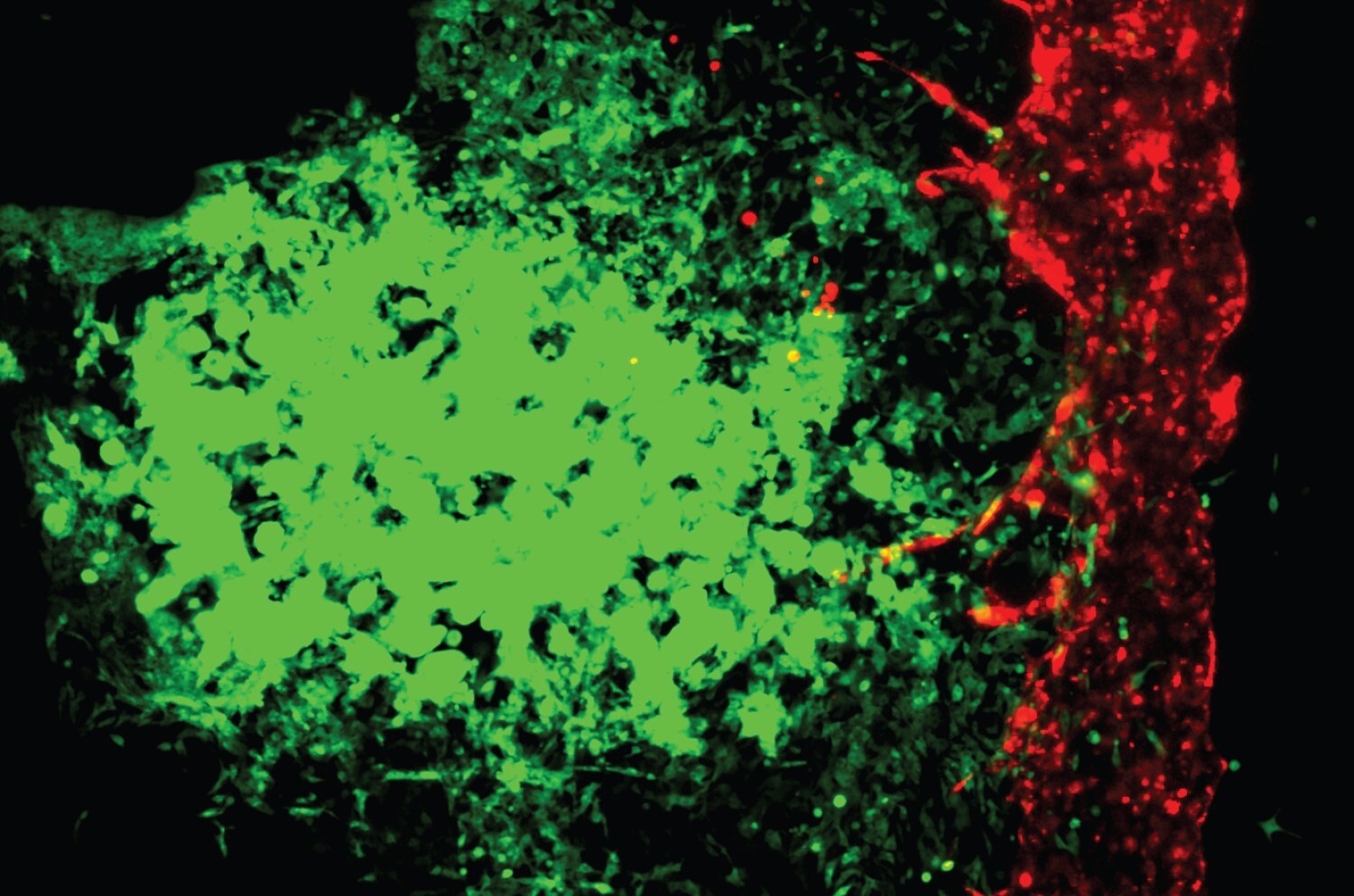
In this facility, where biology and engineering intersect, teams made up of stem cell scientists, biomedical engineers, mechanical engineers, biomaterials scientists, computer scientists, physiologists, surgeons, and more have collaborated on a variety of projects—from creating small beating hearts to making 3D tissue models to more accurately screen the effectiveness of different cancer therapies.
As Panoskaltsis-Mortari, also vice chair for research in the Medical School’s Department of Pediatrics and a Masonic Cancer Center member, moves the 3D Bioprinting Facility to a larger space with new equipment this fall, she’s excited about what’s to come.
Let’s start from the beginning. What exactly is 3D bioprinting?
If 2D printing is simply printing ink on a sheet of paper, imagine a whole pile of sheets and you layer them on top of one another. So basically, 3D printing is creating a 3D object, one layer at a time on top of the previous layer.
3D bioprinting is 3D printing with biological material, with or without live cells. So you are now printing biological tissue.
How could 3D-bioprinted materials be used for medical purposes?
There are some very rudimentary types of tissues in clinical trials now. Today there are 3D-bioprinted corneas as well as tendons and sheets of skin being evaluated in clinical studies.
What other medical uses could you imagine for 3D bioprinting?
One of the most obvious would be 3D bioprinting an organ for transplantation. You would use “ink” made from the patient’s own stem cells, or cells from a stem cell bank that you could match to the patient, so the organ wouldn’t be rejected by the patient’s immune system.
Scientists in other countries are starting to develop the use of bioprinting in vivo, within the patient. So a surgeon uses a handheld 3D bioprinter like a pen, and it extrudes biological material to repair damaged tissue. These studies are now being done in pigs.
You could also use 3D bioprinting to make pills. I envision that in the future—and maybe this is 50 years from now—people would go to a pharmacy and present their list of medications, and the pharmacist could print them all out in combinations that require only one or two pills, rather than the dozens of pills that some people have to take.
There are many other areas that just aren’t tapped into yet.
What current project are you most excited about?
They’re all exciting! I can’t pick. We’re studying things in a way that they’ve never been studied before. Everything we do is developing new knowledge.
Make a gift to support the 3D Bioprinting Facility.


-2.jpg?w=1100)